PRODUCTS
CellO-IF & CellO-M
High-Resolution In Situ Imaging
of Organoids, Spheroids, and Cells
CellO-IF: The Smarter Way to Label
CellO-IF is an all-in-one reagent
that makes immunofluorescence labeling studies easy, simple, and more accurate.
It eliminates harmful workflow steps that cause loss and damage of delicate samples, allowing you to label the entire sample in its growing habitat. This provides more data with better accuracy.
Broad compatibility:
It is fully compatible with various hydrogels and glass-bottom cell culture plates.
Simplified workflow:
You only need your primary and secondary antibodies plus CellO-IF for the experiment.
In Situ Precision:
You can label the entire sample while it remains in its native microenvironment
(even within hydrogels),
resulting in highly specific labeling
without the need for manual specimen transfer.
CellO-M: The Smarter Way to Microscopy
It is all-in-one platform
for streamlined workflows for 2D/3D cell culture and microscopic analysis. It simplifies protocols and ensures optimal sample preservation for high-quality data.
CellO-M provides a controlled, biocompatible environment for the complete cycle of 2D/3D cell cultures; including growing, cryopreservation, labeling, and imaging .
'CellO-IF and CellO-M can be used together or separately.'
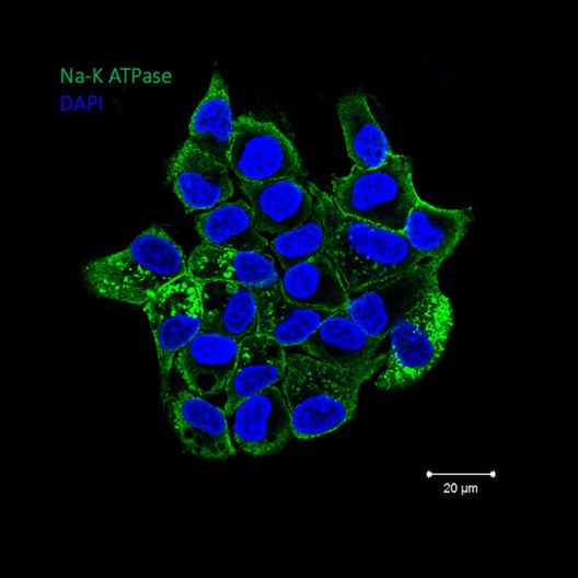
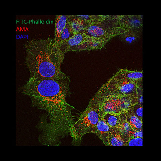
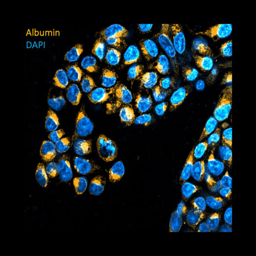
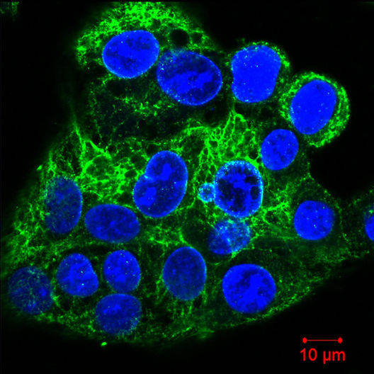
GALLERY 1:
CellO - IF2 in Action
Immunofluorescent Labeling of Cells
GALLERY 2:
CellO - IF3 in Action:
Immunofluorescent Labeling of Organoids and Spheroids
®
GALLERY 3:
CellO-M in Action
Effortless Workflow + High-Quality Data
Culture and analyze your organoids, spheroids, or cells under any microscope—light, confocal, or electron—in a single, contamination-free environment
New Products
*Patent Pending Technologies
Cellorama Technologies:
Your partner in preclinical studies.
Minimize damage, reduce errors, and ensure reliability
with simple, user-friendly, and cost-effective tools.


