top of page

®
Bismillahirrahmanirrahim
Analyze Smarter.
Discover Faster.
We help you improve data quality
in 2D/3D cell culture models by ensuring you capture
every detail of the specimen without losing important information.
Focus on breakthroughs, not sample prep.


Jenny Lai, MD-PhD
Harvard Medical School
“Using CellO-IF simplified our workflow, and it has great potential to make high-throughput imaging of organoids feasible. "
Didem Demirbas, PhD
Boston Children's Hospital
"We really didn’t think imaging the neuronal organoids would be as easy because they are so fragile and difficult to section with traditional methods. We got beautiful images using CellO-IF on both 2D and 3D cultures."
Liyuan Gong, PhD
University at Buffalo, NY
“It indeed helped us savea lot of time staining our organoids.
Overall, the product has been good and helpful for our research.
Thanks for your continuous support!
.”
Ozgecan Kayalar, PhD
Koc University
“We could protect delicate patient-derived organoids and improve the results. It is such an
easy-to-use technology."
Soner Turkuner, PhD
Aysen Yazici, MSc
Gebze Technical University
"The technology is so simple and fast. The results are stunning."
Alp Can, MD
Ferda Topal, MD
School of Medicine
Ankara University
" We achieve consistent results that surpass anything we've done before. The improvement is undeniable!"
Images From Worldwide Users

Lung Organoid / Na-K ATPase

Trophoblast Organoid
Brain Organoid ( Pax2, DAPI)

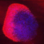
Lung Organoid (Red-Mitochondria, Blue-DAPI))

Liver Cancer Spheroid (Red-Na-K ATPase, Blue-DAPI)

Lung Organoid

Primary Neurons

Hypothalamic Neurons (Green-SIK2, Red-Sytox)

Induced Pluripotent Stem Cells (green – MAP2, red – CREB, blue – DAPI)

bottom of page