®
Bismillahirrahmanirrahim
Analyze Smarter.
Discover Faster.
Standardized 2D & 3D Imaging for Next-Gen Drug Discovery
Empowering studies on stem cells, organoids, spheroids, and organ-on-chips with streamlined workflows and high-quality imaging,
ensuring every critical detail is captured without losing essential data.
CellO-IF
The Smarter Way To Label
Get consistent, high-quality labeling for Organoids, Spheroids, and Cells.
CellO-IF is an all-in-one reagent that simplifies traditional immunofluorescence labeling workflows and eliminates errors.
Instead of spending hours mixing complex buffers and worrying about protocol errors, CellO-IF standardizes everything into eight steps.
It gives you incredible image quality and deep antibody penetration in a fraction of the time, so you can focus on your results.

CellO-M
The Smarter Way To Microscopy
Increase efficiency by culturing and analyzing your entire sample under any microscope—light, confocal, or electron—in a single, contamination-free environment: CellO-M.
Why risk sample loss moving your tissues from plate to plate? CellO-M lets you grow, stain, and analyze cells, organoids and spheroids in their native, optimum environment.
Protect your sample integrity, capture high-fidelity imaging data, and effortlessly transition to high-resolution ultrastructural analysis.

What Users Say
Jenny Lai, MD-PhD
Harvard Medical School
“Using CellO-IF simplified our workflow, and it has great potential to make high-throughput imaging of organoids feasible. "
Didem Demirbas, PhD
Boston Children's Hospital
"We really didn’t think imaging the neuronal organoids would be as easy because they are so fragile and difficult to section with traditional methods. We got beautiful images using CellO-IF on both 2D and 3D cultures."
Liyuan Gong, PhD
University at Buffalo, NY
“It indeed helped us savea lot of time staining our organoids.
Overall, the product has been good and helpful for our research.
Thanks for your continuous support!
.”
Ozgecan Kayalar, PhD
Koc University
“We could protect delicate patient-derived organoids and improve the results. It is such an
easy-to-use technology."
Soner Turkuner, PhD
Aysen Yazici, MSc
Gebze Technical University
"The technology is so simple and fast. The results are stunning."
Alp Can, MD
Ferda Topal, MD
School of Medicine
Ankara University
" We achieve consistent results that surpass anything we've done before. The improvement is undeniable!"
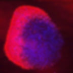
Images From Worldwide Users